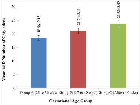
Introduction: The placenta has drawn attention as an important indicator of intrauterine condition of fetus and maternal diseases. The study of number of cotyledons of placenta in different gestational age groups of healthy pregnant mother of Bangladesh is cross sectional descriptive study. Aim of the study: The aim of the study was to study the variation of number of cotyledons of placenta in deferent state of gestational ages of healthy pregnant mother. Methods: This cross sectional descriptive study was conducted in the Department of Anatomy, Mymensingh Medical College, Mymensingh, from January 2018 to December 2018. This study was performed on 80 human placentae. Result: The mean (± SD) number of cotyledon of the placenta was 18.56 (±2.15) in group A, 21.21 (±3.11) in group B and 23.75 (±3.45) in group C. The mean number of cotyledon of the placenta was maximum in group C (23.75) and was minimum in group A (18.56). It was also observed that the mean number of cotyledon of the placenta increased with gestational age. The mean difference of the number of placental cotyledons between groups A and C was statistically moderately significant (p < 0.05) but between A and B and B and C was statistically non-significant (p > 0.05). Conclusion: The placental examination becomes important as it will help in understanding the specific etiologies of adverse outcome. This study has shown that the mean number of cotyledon of the placenta increased with gestational age.
| Published in | American Journal of Health Research (Volume 12, Issue 4) |
| DOI | 10.11648/j.ajhr.20241204.15 |
| Page(s) | 104-109 |
| Creative Commons |
This is an Open Access article, distributed under the terms of the Creative Commons Attribution 4.0 International License (http://creativecommons.org/licenses/by/4.0/), which permits unrestricted use, distribution and reproduction in any medium or format, provided the original work is properly cited. |
| Copyright |
Copyright © The Author(s), 2024. Published by Science Publishing Group |
Placenta, Cotyledons, Gestational Age, Healthy Pregnant Mother
Group | Gestational Age in week | Number of specimen |
|---|---|---|
A | 28 – 36 | 20 |
B | 37 – 40 | 42 |
C | Above 40 weeks | 18 |
Total | 80 |
Gestational Age Group | Number of Specimen (n = 80) | Number of Cotyledons Mean±SD (Minimum – Maximum) |
|---|---|---|
A (28 to 36 weeks) | 18 | 18.56±2.15 (16 – 24) |
B (37 to 40 weeks) | 42 | 21.21±3.11 (17 – 29) |
C (Above 40 weeks) | 20 | 23.75±3.45 (17 – 30) |
Comparison between gestational age groups | Mean Difference | Standard Error of Difference | t | p | Level of significance |
|---|---|---|---|---|---|
A & B | -2.65873 | 0.69781 | -2.01 | 0.05 | Non-significant |
B & C | -2.53571 | 0.90811 | -0.429 | 0.67 | Non-significant |
A & C | 5.19444 | 0.92219 | 3.325 | 0.002 | Moderately significant |
MMCH | Mymensingh Medical College Hospital |
| [1] | Harold Fox, Neil J (2007), Pathology of the placenta, 3rd ed. Philadelphia: Elsevier Saunders. |
| [2] | Udainia A, Bhagwat SS, Mehta CD 2004, ‘Relation between placental surface area, infarction and fetal distress in pregnancy induced hypertension with its clinical relevance’, Journal of Anatomical Society of India, vol. 53, no. 1, pp. 27-30. |
| [3] | Kurdukar MD, Deshpande NM, Shete SS, Zawar MP 2007, ‘Placenta in PIH’, Indian Journal of Pathology and Microbiology, vol. 50, no. 3, pp. 493-497. |
| [4] | Dutta DC 2015, DC Dutta’s textbook of obstetrics including perinatology and contraception, 8th edn, Jaypee Brothers Medical Publishers Pvt. Ltd, New Delhi, pp. 28–45. |
| [5] | Decherney AH, Nathan L, Laufer N, Roman AS 2018, Current diagnosis & treatment obstetrics & gynaecology, 12th edn, McGraw Hill Education, New York, pp. 108, 171–8. |
| [6] | Robertson WB, Brosens I, Dixon HG 1967, ‘The pathological response of the vessels of the placental bed to hypertensive pregnancy’, J. Pathol. Bacteriol. Vol. 93, pp. 581-92. |
| [7] | Benirschke K, Kaufman P, Baergen R 2000, The pathology of the human placenta, 4th edn, Springer, New York, pp. 12–4. |
| [8] | Barker D, Osmond C, Grant S, Thornburg KL, Cooper C, Ring S et al. 2013, ‘Maternal cotyledons at birth predict blood pressure in childhood’, Placenta, Elsevier Ltd. Vol. 34, no. 2013, pp. 672-675. |
| [9] | Kishwara S, Ara S, Rayhan KA, Begum M 2009, ‘Morphological changes of placenta in preeclampsia’, Bangladesh Journal of Anatomy, vol. 7, no. 2, pp. 49–54. |
| [10] | Gunasegaran JP 2017, Textbook of histology, 3rd edn., Reed Elsevier India Pvt. Ltd., New Delhi, pp. 334 – 6. |
| [11] | Sadler TW 2015, Langman’s medical embryology, 13th edn, Wolters Kluwer, Philadelphia, pp. 48, 109–19, 394. |
| [12] | Datta AK 2014, Essentials of human embryology, 6th edn, Current Books International, Kolkata, pp. 21, 35, 57–68. |
| [13] | Ragunath G, Vijayalakshmi Shenoy, V 2011, ‘A study of morphology & morphometry of the human placenta and its clinical relevance in a population in Tamil Nadu’, J. of clinical and diagnostic research, vol. 5, no. 2, pp. 282–6. |
| [14] | Begum T 2010, Gross and histomorphological study of human placenta and umbilical cord in different gestational age group in Bangladesh, thesis, Mymensingh Medical College, Mymensingh. |
| [15] | Majumdar S, Dasgupta H, Bhattacharya K, Bhattacharya A 2005, ‘A study of placenta in normal and hypertensive pregnancies’, Journal of the Anatomical Society of India, vol. 54, no. 2, pp. 1–9. |
| [16] | Sultana S 2005, A comparative study of gross and histomorphological changes of human placenta and umbilical cord in normal and eclamptic pregnancy, thesis, Mymensingh Medical College, Mymensingh. |
| [17] | Dawn CS 2004, Textbook of obstetrics and neonatology, 16th edn, Pratap Medical Publishers, Kolkata, pp. 46–50. |
| [18] | Singh V 2017, Textbook of clinical embryology, 2nd edn, Elsevier Health Sciences, India, pp. 77–8. |
| [19] | Kaufmann P 1985, ‘Basic morphology of the fetal and maternal circuits in the human placenta’, Contrib Gynecol Obstet, vol. 13, no. 1, pp. 5–17. |
| [20] | Boyd JD, Hamilton WJ 1970, The human placenta, 1st edn, Heffer, England, pp. 143, 264. |
APA Style
Bose, S. K., Jabeen, L., Shanto, R. A., Khanam, A., Naznin, R. A., et al. (2024). Study of Number of Cotyledons of Placenta in Different Gestational Age Groups of Healthy Pregnant Mother of Bangladesh. American Journal of Health Research, 12(4), 104-109. https://doi.org/10.11648/j.ajhr.20241204.15
ACS Style
Bose, S. K.; Jabeen, L.; Shanto, R. A.; Khanam, A.; Naznin, R. A., et al. Study of Number of Cotyledons of Placenta in Different Gestational Age Groups of Healthy Pregnant Mother of Bangladesh. Am. J. Health Res. 2024, 12(4), 104-109. doi: 10.11648/j.ajhr.20241204.15
AMA Style
Bose SK, Jabeen L, Shanto RA, Khanam A, Naznin RA, et al. Study of Number of Cotyledons of Placenta in Different Gestational Age Groups of Healthy Pregnant Mother of Bangladesh. Am J Health Res. 2024;12(4):104-109. doi: 10.11648/j.ajhr.20241204.15
@article{10.11648/j.ajhr.20241204.15,
  author = {Sanjib Kumar Bose and Labiba Jabeen and Rafuja Afrin Shanto and Afsana Khanam and Rawshon Ara Naznin and Sharmin Akter Sumi},
  title = {Study of Number of Cotyledons of Placenta in Different Gestational Age Groups of Healthy Pregnant Mother of Bangladesh
},
  journal = {American Journal of Health Research},
  volume = {12},
  number = {4},
  pages = {104-109},
  doi = {10.11648/j.ajhr.20241204.15},
  url = {https://doi.org/10.11648/j.ajhr.20241204.15},
  eprint = {https://article.sciencepublishinggroup.com/pdf/10.11648.j.ajhr.20241204.15},
  abstract = {Introduction: The placenta has drawn attention as an important indicator of intrauterine condition of fetus and maternal diseases. The study of number of cotyledons of placenta in different gestational age groups of healthy pregnant mother of Bangladesh is cross sectional descriptive study. Aim of the study: The aim of the study was to study the variation of number of cotyledons of placenta in deferent state of gestational ages of healthy pregnant mother. Methods: This cross sectional descriptive study was conducted in the Department of Anatomy, Mymensingh Medical College, Mymensingh, from January 2018 to December 2018. This study was performed on 80 human placentae. Result: The mean (± SD) number of cotyledon of the placenta was 18.56 (±2.15) in group A, 21.21 (±3.11) in group B and 23.75 (±3.45) in group C. The mean number of cotyledon of the placenta was maximum in group C (23.75) and was minimum in group A (18.56). It was also observed that the mean number of cotyledon of the placenta increased with gestational age. The mean difference of the number of placental cotyledons between groups A and C was statistically moderately significant (p  0.05). Conclusion: The placental examination becomes important as it will help in understanding the specific etiologies of adverse outcome. This study has shown that the mean number of cotyledon of the placenta increased with gestational age.
},
 year = {2024}
}
TY  - JOUR
T1  - Study of Number of Cotyledons of Placenta in Different Gestational Age Groups of Healthy Pregnant Mother of Bangladesh

AU  - Sanjib Kumar Bose
AU  - Labiba Jabeen
AU  - Rafuja Afrin Shanto
AU  - Afsana Khanam
AU  - Rawshon Ara Naznin
AU  - Sharmin Akter Sumi
Y1  - 2024/08/30
PY  - 2024
N1  - https://doi.org/10.11648/j.ajhr.20241204.15
DO  - 10.11648/j.ajhr.20241204.15
T2  - American Journal of Health Research
JF  - American Journal of Health Research
JO  - American Journal of Health Research
SP  - 104
EP  - 109
PB  - Science Publishing Group
SN  - 2330-8796
UR  - https://doi.org/10.11648/j.ajhr.20241204.15
AB  - Introduction: The placenta has drawn attention as an important indicator of intrauterine condition of fetus and maternal diseases. The study of number of cotyledons of placenta in different gestational age groups of healthy pregnant mother of Bangladesh is cross sectional descriptive study. Aim of the study: The aim of the study was to study the variation of number of cotyledons of placenta in deferent state of gestational ages of healthy pregnant mother. Methods: This cross sectional descriptive study was conducted in the Department of Anatomy, Mymensingh Medical College, Mymensingh, from January 2018 to December 2018. This study was performed on 80 human placentae. Result: The mean (± SD) number of cotyledon of the placenta was 18.56 (±2.15) in group A, 21.21 (±3.11) in group B and 23.75 (±3.45) in group C. The mean number of cotyledon of the placenta was maximum in group C (23.75) and was minimum in group A (18.56). It was also observed that the mean number of cotyledon of the placenta increased with gestational age. The mean difference of the number of placental cotyledons between groups A and C was statistically moderately significant (p  0.05). Conclusion: The placental examination becomes important as it will help in understanding the specific etiologies of adverse outcome. This study has shown that the mean number of cotyledon of the placenta increased with gestational age.

VL  - 12
IS  - 4
ER  -
Department of Anatomy, Gonoshasthaya Samaj Vittik Medical College, Dhaka, Bangladesh
Department of Anatomy, Asgar Ali Medical College, Dhaka, Bangladesh
Department of Anatomy, Dhaka Central International Medical College, Dhaka, Bangladesh
Department of Anatomy, TMSS Medical College, Bogura, Bangladesh
Department of Anatomy, TMSS Medical College, Bogura, Bangladesh
Department of Anatomy, Bangabandhu Sheikh Mujib Medical University (BSMMU), Dhaka, Bangladesh